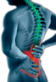
Background on Sacroiliac Joint Dysfunction
For decades, the sacroiliac joint was suspected to be a common cause of low back and/or leg pain, although difficulty in proving it with standard diagnostic tests left many in the medical profession skeptical.
Also, over the last twenty to thirty years, the medical profession has focused more on discogenic pain (herniated disc, degenerative disc disease) as a common cause of low back and/or leg pain. In fact, to this day sacroiliac joint dysfunction remains difficult to diagnose, but anesthetic injection blocks specifically applied to the SI joint are considered the gold standard.
Accurately diagnosing sacroiliac joint pain can be difficult because the symptoms mimic other common conditions, including other mechanical back pain conditions like facet syndrome as well as other lumbar spine conditions including disc herniation and radiculopathy (pain along the sciatic nerve that radiates down the leg).
A diagnosis is usually arrived at through physical examination (eliminating other causes) and/or an injection (utilized to block the pain).
Physical Examination to Determine the Source of Sacroiliac Pain
In physical examination, the doctor may try to determine if the sacroiliac joint is the cause of pain through movement of the joint. If the movement recreates the patient’s pain, and no other cause of pain can explain the patient’s pain and symptoms (such as a disc herniation on an MRI scan), the sacroiliac joint may be the cause of the pain.
There are several orthopedic provocative tests that can be used in an attempt to reproduce the symptoms associated with sacroiliac joint pain. As a rule, several positive tests that reproduce pain specifically located at the sacroiliac joint improves the probability of the diagnosis of sacroiliac joint dysfunction.
For example, a physical exam may consist of the following:
- The patient may lie face up on the edge of an examination table with the leg of the affected side hanging off the side of the table towards the floor or resting on a nearby stool so that the sacroiliac joint lies on the edge of the table.
- The opposite knee can be drawn to the chest to further isolate the SI joint on the side of the hanging leg. In this position, there is no support for the hip joint and pressing down on the iliac crest (pelvis) or upper thigh may reproduce the patient’s pain.
- Care in locating the pain directly over the SI joint is needed as other structures of the lumbar spine and pelvis are also stressed in this position.
Injections to Determine the Source of Pain
A sacroiliac joint injection, sometimes called a sacroiliac joint block, can be a useful diagnostic test. It takes a highly skilled and experienced physician to be able to insert a needle into the correct portion of the sacroiliac joint. Because of this, the injection is usually guided by X-ray to make sure the joint is properly injected. Sometimes a dye is injected so that the joint is better visualized, which is called an arthrogram.
In this test, a physician uses fluoroscopic guidance (live X-ray) and inserts a needle into the sacroiliac joint to inject lidocaine (a numbing solution). If the injection relieves the patient’s pain, it can be inferred that the sacroiliac joint is the source of the pain. Usually, a steroid solution is injected at the same time to decrease inflammation in the sacroiliac joint and decrease pain.