Facet joint injections involve an injection of anti-inflammatory steroid solution directly into the joint. If such an injection confirms the facet joint as the likely source of the patient’s pain, but this injection – along with other treatments (such as physical therapy, manual manipulation, and medications) have not resulted in long term pain relief, then a medial branch block may be recommended.
As evidence evolves on the efficacy of facet joint injections, a medial branch block may also be considered instead of a facet joint injection. A medial branch block might also be considered first if for any reason the patient cannot tolerate the steroid and/or an injection directly into the facet joint.
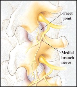
For the facet joints, the medial branch nerves are the small nerves that carry pain signals to the brain.
Facet joints are pairs of small joints that are situated at each vertebral level of the spine. A facet joint may also be called a zygapophysial joint or a Z-joint.
Medial Nerves
Each facet joint is connected to two medial nerves that carry pain signals away from the spine to the brain.
The medial nerves are uniquely located in each segment of the spine:
- Cervical medial branch nerves are located in a bony groove in the neck
- Thoracic medial branch nerves are located over a bone in the mid-back or upper back
- Lumbosacral medial branch nerves are found in a bony groove in the low back
Medial Nerve Function
The medial branch nerve block is designed to interrupt the nerves ability to transmit the pain signals to the brain, which in turn will determine – or diagnose – if the facet joint(s) is the source of the patient’s pain.
These medial branch nerves do not control any major muscles or carry any sensation in the arms or legs, so there is no danger of negatively affecting those areas – or negatively affecting other pain sensing processes – with a medial branch block.
The medial branch nerves do control small muscles in the neck, the mid and lower back, but loss of these nerves has not proved harmful.
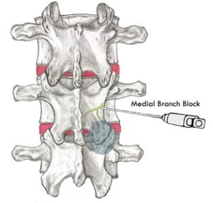
Medial Branch Block Procedure
As with many spinal injections, medial branch nerve block procedures are best performed under fluoroscopy (live X-ray) for guidance in properly targeting the nerves, placing the needle, and avoiding injury.
On the day of the injection, patients are advised to avoid driving and doing any strenuous activities, and to get plenty of rest the night before.
Medial Branch Nerve Block Steps
The medial branch injection procedure includes the following steps:
- Commonly, the procedure is performed without any sedation, however, an IV line can be started if relaxation medicine is needed
- The patient lies face down on an procedure table, and the skin over the area to be tested is well cleansed
- The physician treats a small area of skin with a numbing medicine (anesthetic), which may sting for a few seconds
- The physician uses X-ray guidance (fluoroscopy) to direct a very small needle over the medial branch nerves
- A small amount of contrast dye is then injected to confirm that the medicine covers the medial branch nerve
- Following this confirmation, a small amount of numbing medicine (anesthetic) will then be slowly injected onto each targeted nerve.
The injection itself only takes a few minutes, but the entire procedure usually takes between fifteen and thirty minutes.
After the Injection
After the procedure, the patient typically remains resting in the recovery area for 20 to 30 minutes. The physician will then ask the patient to perform some movements or activities that would usually provoke the pain. This assessment is done in order to determine if the medial branch nerve block has reduced the patient’s pain.